


Introduction
The human brain is constantly producing electrical activity as billions of nerve cells communicate with one another. EEG tech, short for electroencephalography technology, gives clinicians and researchers a practical way to record patterns associated with that activity from electrodes placed on or near the head.
An electroencephalogram, commonly called an EEG, records electrical activity using electrodes, most commonly positioned on the scalp. The resulting signals appear as changing waveforms that trained professionals can evaluate for patterns associated with different states and neurological conditions. citeturn0search0turn0search2
Although EEG has been used in medicine for decades, the technology continues to evolve. Modern systems may incorporate digital signal processing, wireless hardware, portable equipment, quantitative analysis software, and applications extending from hospital monitoring to neuroscience research and brain-computer interfaces.
This guide explains what EEG tech is, how it works, its major components and types, where it is used, and the advantages and limitations that users should understand.
Table of Contents
- What Is EEG Tech?
- How Does EEG Technology Work?
- Understanding EEG Brain Waves
- Key Components of an EEG System
- Types of EEG Technology
- Key Features of Modern EEG Tech
- Major Uses of EEG Technology
- Benefits of EEG Technology
- Pros and Cons of EEG Tech
- EEG Technology vs Other Brain-Imaging Methods
- The Future of EEG Technology
- Frequently Asked Questions
- Conclusion
- Key Takeaways
What Is EEG Tech?
EEG tech refers to the hardware, electrodes, software, and signal-processing methods used to acquire and analyze electrical signals associated with brain activity.
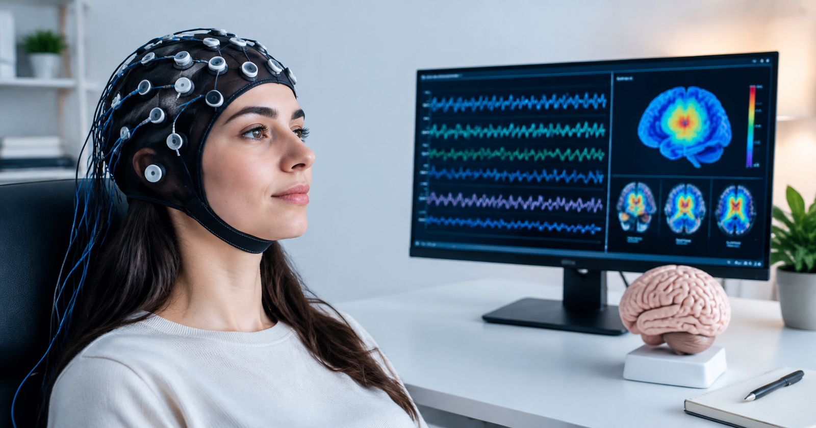
Traditional clinical EEG systems use electrodes positioned at defined locations across the scalp. The electrodes detect extremely small voltage differences generated by the combined electrical activity of groups of neurons.
Importantly, standard scalp EEG electrodes are recording sensors. They do not normally send electricity into the brain. This is one reason routine EEG testing is considered noninvasive and generally painless. citeturn0search0
Modern EEG equipment can range from conventional wired clinical machines to portable and wireless systems. The FDA, for example, distinguishes between reduced-montage systems with fewer than 16 electrodes and full-montage systems using 16 or more electrodes. citeturn0search3turn0search4
How Does EEG Technology Work?
To understand EEG technology, it helps to follow the signal from the brain to the computer screen.
1. Brain Cells Produce Electrical Activity
Neurons communicate through electrochemical processes. When large populations of neurons become active in coordinated patterns, their combined electrical activity can produce voltage changes detectable at the scalp.
EEG does not normally measure the electrical activity of a single neuron. Instead, it records the summed activity produced by populations of neurons, particularly activity generated near the cerebral cortex.
2. Electrodes Detect Voltage Differences
Small electrodes are placed at specific locations on the scalp. Depending on the system, they may be individual metal discs attached with conductive material or incorporated into an EEG cap.
These electrodes detect voltage differences between recording locations.
Electrode placement matters because activity recorded over one part of the scalp can differ from activity recorded elsewhere. Standardized placement helps clinicians compare signals across different brain regions and recordings.
3. The Signal Is Amplified
Brain signals recorded at the scalp are extremely small. EEG hardware therefore uses amplifiers to increase signal strength so the electrical activity can be processed and displayed.
Modern EEG amplifiers are designed to capture subtle biological signals while minimizing interference.
4. Analog Signals Are Converted Into Digital Data
After amplification, the electrical signals are converted into digital information.
The computer can then store, process, filter, visualize, and analyze the recording. Modern electroencephalographs commonly include recording hardware, computers, monitors, data storage, and software for displaying and annotating EEG signals. citeturn0search4
5. Software Displays the EEG
EEG software typically displays the recorded signals as multiple waveform channels.
A trained clinician can examine factors such as:
- Frequency
- Amplitude
- Symmetry
- Timing
- Location
- Background rhythms
- Response to stimulation
- Abnormal discharges
Software may also perform mathematical analysis. Quantitative EEG tools, for example, can calculate parameters such as total power, frequency-related measurements, and asymmetry. citeturn0search14
6. Specialists Interpret the Recording
EEG technology collects the information, but clinical interpretation requires expertise and appropriate context.
An EEG result is not simply classified as “good” or “bad.” Clinicians consider the patient’s symptoms, medical history, medications, age, state of wakefulness, and other diagnostic findings when interpreting a recording.
Understanding EEG Brain Waves
EEG recordings contain electrical rhythms at different frequencies. These frequency ranges are commonly described using names such as delta, theta, alpha, beta, and gamma.
Delta Waves
Delta activity generally refers to very slow EEG frequencies and is prominent during deep sleep.
The significance of slow activity depends strongly on factors such as age, state of consciousness, distribution, and clinical circumstances.
Theta Waves
Theta rhythms are faster than delta waves and may appear during drowsiness and certain stages of sleep.
They can also occur in waking recordings, with their significance depending on age and context.
Alpha Waves
Alpha activity is commonly observed in relaxed, awake adults, particularly when the eyes are closed.
It is often most prominent over posterior areas of the head and may change when the person opens their eyes or becomes mentally engaged.
Beta Waves
Beta activity consists of relatively faster frequencies and can be associated with alert wakefulness.
Certain medications and muscle activity can also influence faster frequencies observed during EEG recordings.
Gamma Activity
Gamma refers to higher-frequency activity that has attracted considerable interest in neuroscience research.
However, high-frequency scalp recordings require careful interpretation because muscle activity and other sources can contaminate the signal.
Key Components of an EEG System
A modern EEG setup involves considerably more than electrodes.
EEG Electrodes
Electrodes provide the interface between the recording system and the subject. They are positioned according to the requirements of the examination and the type of EEG equipment being used.
Clinical systems may use individual electrodes or electrode caps designed to simplify placement.
Conductive Gel or Paste
Conductive materials can improve electrical contact between an electrode and the scalp.
Good electrode contact helps reduce unwanted noise and improves recording quality.
EEG Amplifier
The amplifier increases the strength of the tiny biological signals detected by the electrodes.
High-quality amplification is essential because EEG signals can easily be affected by environmental electrical noise.
Analog-to-Digital Converter
This component converts recorded analog voltage changes into digital information that computers can process.
Sampling rate and signal resolution are important technical considerations because they influence how accurately the original waveform is represented.
EEG Software
Software allows professionals to display, store, annotate, review, and sometimes analyze EEG recordings.
Some specialized applications provide quantitative analysis, while others may support source localization or other advanced processing. FDA classifications distinguish standard recording software from more complex analytical applications. citeturn0search10turn0search11
Computer and Data Storage
Digital EEG recordings can contain substantial amounts of data, particularly during continuous or long-term monitoring.
Computers are therefore used for recording, visualization, storage, retrieval, and analysis.
Types of EEG Technology
EEG tech is available in several forms because different applications require different recording durations, electrode arrangements, and levels of mobility.
Routine EEG
A routine EEG is one of the most familiar forms of the test.
Electrodes are placed on the scalp while brain activity is recorded for a limited period. A typical routine recording may take roughly 20 to 40 minutes once the electrodes are in place, although the complete appointment may be longer. citeturn0search0
During the examination, the patient may be asked to open or close their eyes, breathe deeply, or look at flashing lights.
Sleep EEG
Some abnormalities become easier to detect during sleep.
A clinician may therefore request an EEG that includes natural or induced sleep. Patients can sometimes be asked to reduce their sleep beforehand so they are more likely to fall asleep during the recording.
Ambulatory EEG
Ambulatory EEG allows brain activity to be recorded for longer periods while a person is outside the traditional EEG laboratory.
Recording over one or more days can increase the chance of capturing intermittent events that might not occur during a short routine examination. citeturn0search0
Video EEG Monitoring
Video EEG combines synchronized video with the EEG recording.
This allows clinicians to compare physical behavior with electrical brain activity. It can be particularly valuable when specialists need to determine whether an observed episode corresponds with a particular EEG pattern.
Continuous EEG
Continuous EEG monitoring may be used in hospitals and intensive care environments when brain activity needs to be observed for an extended period.
For example, it can help clinicians detect ongoing or recurrent seizure activity in critically ill patients who may not show obvious physical signs.
Quantitative EEG
Quantitative EEG, often abbreviated qEEG, applies mathematical processing to recorded EEG data.
Software can calculate conventional parameters such as spectral power, frequency-related measurements, and asymmetry. These outputs must still be interpreted appropriately rather than treated as automatic diagnoses. citeturn0search14
High-Density EEG
High-density EEG uses a larger number of electrodes distributed across the scalp.
More recording locations can provide improved spatial sampling, making this approach valuable for certain research and specialized clinical applications.
Sub-Scalp EEG
Some newer systems use electrodes positioned beneath the scalp without penetrating brain tissue.
The FDA describes sub-scalp implanted EEG systems intended for long-term acquisition, transmission, and storage of brain electrical activity for remote monitoring of patients with epilepsy. citeturn0search5
Key Features of Modern EEG Tech
Modern EEG systems can offer features that were difficult to achieve with older equipment.
High temporal resolution: EEG can capture very rapid changes in electrical brain activity, making it particularly useful when timing is important.
Digital recording: Signals can be stored electronically for later review, annotation, processing, and comparison.
Multi-channel recording: Multiple electrodes allow simultaneous observation of activity across different scalp locations.
Long-term monitoring: Ambulatory and continuous systems can record activity for hours or days.
Video synchronization: EEG waveforms can be synchronized with video to correlate electrical activity with physical events.
Wireless capabilities: Some contemporary EEG hardware can incorporate wireless components, reducing the restrictions associated with traditional wired configurations. citeturn0search4
Software-based analysis: Digital processing can calculate EEG parameters and assist qualified professionals with reviewing large datasets.
Major Uses of EEG Technology
Diagnosing and Managing Epilepsy
Epilepsy evaluation remains one of the best-known uses of EEG technology.
Certain EEG patterns can support the diagnosis and classification of seizure disorders. EEG testing may also provide information relevant to assessing the likelihood of further seizures. citeturn0search0turn0search17
However, EEG findings must be interpreted alongside the clinical history because a normal routine EEG does not automatically exclude epilepsy.
Investigating Unexplained Episodes
EEG may be requested when a person experiences unexplained periods of altered awareness, unusual movements, collapse, or other events that could potentially involve abnormal brain activity.
Longer-term or video EEG can be particularly useful when capturing the event itself is important.
Sleep Assessment
EEG is an important part of studying sleep because different stages of sleep produce recognizable changes in brain activity.
In comprehensive sleep studies, EEG data may be recorded alongside breathing, eye movements, heart activity, muscle activity, and oxygen measurements.
Intensive Care Monitoring
Continuous EEG can help clinicians monitor neurological function in critically ill patients.
It may be especially important for detecting electrical seizures that have few or no visible physical signs.
Evaluating Brain Disorders
Changes in EEG activity can provide useful information in the evaluation of several neurological conditions.
Clinical sources describe applications involving encephalopathy, brain injury, inflammation of the brain, stroke, coma, and other disorders affecting brain function. citeturn0search0turn0search2
An EEG is generally one part of a broader diagnostic assessment rather than a standalone answer.
Neuroscience Research
Researchers use EEG technology to investigate how brain activity changes during perception, attention, memory, decision-making, language, movement, and sleep.
Its excellent temporal resolution makes EEG especially useful when researchers need to understand when a neural response occurs.
Brain-Computer Interfaces
A brain-computer interface, or BCI, attempts to translate measurable brain activity into commands for an external system.
EEG is attractive for BCI research because scalp recordings can be obtained without surgery. Experimental systems have explored applications such as communication interfaces, assistive technologies, rehabilitation tools, and computer control.
Neurofeedback
Neurofeedback uses measurements of brain activity to provide feedback to a participant, often in real time.
EEG-based neurofeedback remains an active area of clinical and scientific research. Evidence and appropriate indications vary by condition, so it should not be presented as a universal treatment.
Benefits of EEG Technology
One of the greatest advantages of EEG tech is its ability to measure changes in brain electrical activity with excellent timing precision.
It is also generally noninvasive when scalp electrodes are used. Routine EEG is considered safe and painless because the electrodes record signals rather than delivering electrical stimulation. citeturn0search0
EEG equipment can also be more portable than large imaging technologies. Ambulatory systems make it possible to record patients for extended periods outside a traditional hospital laboratory.
Another advantage is flexibility. The same fundamental technology can support routine neurological testing, long-term monitoring, sleep-related assessment, neuroscience research, and emerging human-computer interaction studies.
Pros and Cons of EEG Tech
Pros
- Excellent temporal resolution
- Standard scalp EEG is noninvasive
- Generally painless
- Useful for detecting abnormal electrical activity
- Particularly important in seizure and epilepsy evaluation
- Can support prolonged monitoring
- Portable and ambulatory systems are available
- Can be synchronized with video
- Valuable for both clinical care and research
- Typically does not expose patients to ionizing radiation
Cons
- Scalp EEG has limited spatial resolution compared with some imaging techniques
- Signals can be affected by eye movements and muscle activity
- Electrical interference can reduce recording quality
- Hair and poor electrode contact may create technical challenges
- A short recording can miss intermittent abnormalities
- Interpretation requires specialized expertise
- Normal EEG findings do not necessarily rule out neurological disease
- EEG usually needs to be considered alongside clinical findings and other tests
EEG Technology vs Other Brain-Imaging Methods
EEG is sometimes grouped with MRI, CT, PET, and MEG, but these technologies measure different things.
EEG primarily records electrical activity. MRI provides detailed structural images, while functional MRI estimates changes associated with brain activity through blood-oxygen-related signals.
CT uses X-rays to create structural images and can be especially useful in urgent medical situations. PET uses radioactive tracers to examine physiological or metabolic processes.
EEG’s major strength is temporal resolution. It can follow electrical changes occurring over very short time intervals.
Its weakness is determining the precise anatomical origin of those signals. Electrical activity spreads through brain tissue, cerebrospinal fluid, the skull, and scalp before reaching surface electrodes.
For this reason, EEG and imaging technologies are often complementary rather than direct competitors.
The Future of EEG Technology
EEG technology is moving toward smaller hardware, longer monitoring periods, improved connectivity, and more sophisticated software.
Wearable EEG
Portable headsets and compact electrode systems are making EEG easier to use outside specialized laboratories.
Future wearable systems could expand long-term neurological monitoring, rehabilitation research, sleep research, and human-computer interaction.
Remote Monitoring
Long-duration recording is becoming increasingly important, particularly for conditions involving intermittent events.
Sub-scalp systems represent one approach to obtaining extended recordings. FDA-classified systems already exist for continuously acquiring, transmitting, and storing electrical brain activity for remote monitoring in people with epilepsy. citeturn0search5
Artificial Intelligence and EEG Analysis
Machine learning can help researchers process large EEG datasets and identify complex signal patterns.
However, automated analysis requires careful validation. Clinical decisions should not be based simply on an algorithm’s output without considering its intended use, performance, limitations, and professional interpretation.
Better Source Localization
Researchers continue to develop methods for estimating where recorded electrical activity originated within the brain.
Advanced EEG software can combine electrical recordings with neuroimaging information for source localization. The FDA recognizes software specifically designed to correlate EEG or MEG activity with neuroimaging data for this purpose. citeturn0search11
Brain-Computer Interaction
BCI development could become one of the most visible nontraditional applications of EEG technology.
Research continues into communication systems, assistive devices, rehabilitation, adaptive computing, and hands-free interfaces based on measurable neural activity.
Frequently Asked Questions
1. What does EEG tech measure?
EEG tech records voltage changes associated with electrical activity generated by populations of brain cells. In standard EEG testing, electrodes placed on the scalp detect these signals and send them to equipment that amplifies, digitizes, and displays the recording. citeturn0search0
2. Does an EEG send electricity into your brain?
A standard diagnostic scalp EEG does not send electricity into the brain. Its electrodes act as sensors that record naturally occurring electrical activity.
3. What is EEG technology mainly used for?
One of its most established uses is evaluating epilepsy and seizure disorders. EEG can also contribute to assessments involving sleep conditions, brain injury, encephalopathy, coma, and other neurological situations. citeturn0search0turn0search2
4. Is EEG technology safe?
Routine scalp EEG is generally considered safe and painless. In some epilepsy evaluations, procedures such as flashing lights or deep breathing may intentionally increase the chance of provoking abnormal activity, so testing is performed with appropriate clinical supervision. citeturn0search0
5. How long does an EEG take?
A routine EEG recording commonly takes around 20 to 40 minutes after electrode placement. Sleep, ambulatory, video, and continuous EEG monitoring can last considerably longer depending on the clinical objective. citeturn0search0
6. Can an EEG detect epilepsy?
EEG is one of the main tests used to support the diagnosis of epilepsy because it can reveal electrical patterns associated with seizure disorders. However, a normal EEG does not by itself rule out epilepsy, and diagnosis requires clinical evaluation. citeturn0search0turn0search17
7. What is the difference between EEG and MRI?
EEG records electrical brain activity, whereas MRI primarily produces detailed images of brain anatomy. EEG provides excellent information about the timing of electrical events, while MRI generally offers much greater anatomical detail.
8. Can EEG technology read thoughts?
Not in the science-fiction sense. EEG can detect patterns associated with certain brain states or tasks, and controlled BCI systems can sometimes classify predefined patterns. It cannot simply translate a person’s unrestricted private thoughts into complete sentences.
Conclusion
EEG tech provides a direct and practical way to observe electrical brain activity over time. By detecting tiny voltage changes through electrodes and transforming them into interpretable digital recordings, EEG technology gives clinicians and researchers valuable information about how the brain is functioning.
Its established role in epilepsy and seizure evaluation remains particularly important, but modern applications extend much further. Sleep research, intensive care monitoring, neuroscience, quantitative analysis, wearable devices, remote monitoring, and brain-computer interfaces are all contributing to the continued development of EEG.
At the same time, EEG has clear limitations. Scalp recordings can be affected by artifacts, precise source localization can be challenging, and recordings must be interpreted in their clinical context. For these reasons, EEG works best as a specialized measurement tool used alongside professional expertise and, where necessary, other diagnostic technologies.
Read more: Airwave Technologies HSD-Titanium Headset Microphone: Common User Problems and How It Solves Them